



Thrall’s Textbook of Veterinary Diagnostic Radiology, 8th Edition
**Selected for 2025 Doody’s Core Titles® in Veterinary Medicine**
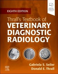
Improve your radiographic interpretation skills, regardless of your level of experience with Textbook of Veterinary Diagnostic Radiology, 8th Edition, your one-stop resource for understanding the principles of radiographic technique and interpretation for dogs, cats, and horses. Within this bestselling text, high-quality radiographic images accompany clear coverage of diagnostic radiology, ultrasound, MRI, and CT. User-friendly direction helps you develop essential skills in patient positioning, radiographic technique and safety measures, normal and abnormal anatomy, radiographic viewing and interpretation, and alternative imaging modalities. This edition has been thoroughly revised to include the latest advances in the field, expand the number of image examples, and include a new ebook with every new print purchase!
Improve your radiographic interpretation skills, regardless of your level of experience with Textbook of Veterinary Diagnostic Radiology, 8th Edition, your one-stop resource for understanding the principles of radiographic technique and interpretation for dogs, cats, and horses. Within this bestselling text, high-quality radiographic images accompany clear coverage of diagnostic radiology, ultrasound, MRI, and CT. User-friendly direction helps you develop essential skills in patient positioning, radiographic technique and safety measures, normal and abnormal anatomy, radiographic viewing and interpretation, and alternative imaging modalities. This edition has been thoroughly revised to include the latest advances in the field, expand the number of image examples, and include a new ebook with every new print purchase!
ISBN :
9780323932356
Publication Date :
16-10-2024
Stock Status :
Please allow 3-4 weeks for delivery
**Selected for 2025 Doody’s Core Titles® in Veterinary Medicine**
Improve your radiographic interpretation skills, regardless of your level of experience with Textbook of Veterinary Diagnostic Radiology, 8th Edition, your one-stop resource for understanding the principles of radiographic technique and interpretation for dogs, cats, and horses. Within this bestselling text, high-quality radiographic images accompany clear coverage of diagnostic radiology, ultrasound, MRI, and CT. User-friendly direction helps you develop essential skills in patient positioning, radiographic technique and safety measures, normal and abnormal anatomy, radiographic viewing and interpretation, and alternative imaging modalities. This edition has been thoroughly revised to include the latest advances in the field, expand the number of image examples, and include a new ebook with every new print purchase!
Improve your radiographic interpretation skills, regardless of your level of experience with Textbook of Veterinary Diagnostic Radiology, 8th Edition, your one-stop resource for understanding the principles of radiographic technique and interpretation for dogs, cats, and horses. Within this bestselling text, high-quality radiographic images accompany clear coverage of diagnostic radiology, ultrasound, MRI, and CT. User-friendly direction helps you develop essential skills in patient positioning, radiographic technique and safety measures, normal and abnormal anatomy, radiographic viewing and interpretation, and alternative imaging modalities. This edition has been thoroughly revised to include the latest advances in the field, expand the number of image examples, and include a new ebook with every new print purchase!
Key Features
- UPDATED! User-friendly content helps you develop essential skills in patient positioning, radiographic technique and safety measures, normal and abnormal anatomy, radiographic viewing and interpretation, and alternative imaging modalities.
- NEW! The latest digital imaging information helps you stay up to date with the latest advances in the field.
- NEW! An ebook version, included with every new print purchase, provides access to all the text, figures, and references, with the ability to search, customize content, make notes and highlights, and have content read aloud. Also included are videos, quizzes, and additional image examples of the most common diseases.
- UPDATED! Current coverage of the principles of radiographic technique and interpretation for the most seen species in private veterinary practices and veterinary teaching hospitals includes the cat, dog, and horse.
- Coverage of special imaging procedures such as the esophagram, upper GI examination, excretory urography, and cystography, helps in determining when and how these procedures are performed in today’s practice.
- Content on abdominal ultrasound imaging helps in deciding on a diagnostic plan and interpreting common ultrasound findings.
- An atlas of normal radiographic anatomy in each section makes it easier to recognize abnormal radiographic findings.
- High-quality radiographic images clarify key concepts and interpretation principles.
Author Information
Edited by Gabriela Seiler, Dr.Med.Vet., DECVDI, DACVR, Professor of Radiology, Molecular Biomedical Sciences, North Carolina State University, Raleigh, North Carolina and Donald E. Thrall, DVM, PhD, Emeritus Professor, College of Veterinary Medicine, North Carolina State University, Raleigh, North Carolina, Radiologist, IDEXX Telemedicine Consultants, Clackamas, Oregon
