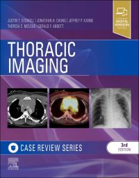
Master the critical imaging content you need to know with this thoroughly updated title in the popular Case Review series. Thoracic Imaging offers a highly illustrated, case-based preparation for board review to help residents and
...view more
Master the critical imaging content you need to know with this thoroughly updated title in the popular Case Review series. Thoracic Imaging offers a highly illustrated, case-based preparation for board review to help residents and recertifying radiologists succeed on exams, demonstrate a clinical understanding of thoracic imaging, and improve imaging accuracy and interpretation. Cases include both common and difficult-to-diagnose disorders spanning the range of diseases impacting the thorax, including sarcoidosis, lymphoma, traumatic aortic injury, small airway disease, idiopathic interstitial pneumonias, developmental lung disease, and lung cancer.
Master the critical imaging content you need to know with this thoroughly updated title in the popular Case Review series. Thoracic Imaging offers a highly illustrated, case-based preparation for board review to help residents and recertifying radiologists succeed on exams, demonstrate a clinical understanding of thoracic imaging, and improve imaging accuracy and interpretation. Cases include both common and difficult-to-diagnose disorders spanning the range of diseases impacting the thorax, including sarcoidosis, lymphoma, traumatic aortic injury, small airway disease, idiopathic interstitial pneumonias, developmental lung disease, and lung cancer.
New to this edition
Clearly see and interpret 400 high-quality, state-of-the-art images representing a wide range of clinical situations encountered in thoracic imaging.
Get fresh perspectives from 200 updated or new cases reflecting the most recent changes in thoracic imaging, including PET-CT, emerging pulmonary infections, smoking-related lung diseases, Fleischner guidelines for management of incidentally detected lung nodules, and ground glass and part-solid lung nodules.
Spend less time searching and more time learning with easy-to-navigate chapters focused on visual identification and diagnosis, and reorganized by degree of case difficulty.
Key Features
Presents more than 145 high-yield case studies organized by level of difficulty, helping you build your knowledge and confidence in stages. All cases have been refreshed and rewritten to capture the latest clinical implications and diagnostic pearls on thoracic conditions that you will be tested on.
Includes multiple-choice questions, answers, and rationales that mimic the format of certification exams.
Uses short, easily digestible chapters covering the full range of thoracic imaging for efficient, effective learning and exam preparation.
Features hundreds of high-quality, full-color images (many new to this edition) representing a wide range of clinical situations encountered in thoracic imaging. Images include chest radiographs, CT, PET-CT, and MRI to help you expand your visual identification and diagnostic skills.
Author Information
By Justin T. Stowell, MD, Department of Radiology, Mayo Clinic, Jacksonville, Florida; Jonathan H. Chung, MD, Section Chief, Thoracic Radiology, Associate Professor, The University of Chicago Medicine, Chicago, Illinois, USA; Jeffrey P. Kanne, MD,, Professor and Chief of Thoracic Imaging, Vice Chair of Quality and Safety, Department of Radiology, University of Wisconsin School of Medicine and Public Health, Madison Wisconsin, USA; Theresa C. McLoud, MD, Associate Radiologist-in-Chief, Massachusetts General Hospital; Director of Education & Professor of Radiology, Harvard Medical School, Boston, MA, USA and Gerald F. Abbott, MD, Associate Radiologist, Thoracic Imaging and Intervention Division, Massachusetts General Hospital, Boston, Massachusetts
By Justin T. Stowell, MD, Department of Radiology, Mayo Clinic, Jacksonville, Florida; Jonathan H. Chung, MD, Section Chief, Thoracic Radiology, Associate Professor, The University of Chicago Medicine, Chicago, Illinois, USA; Jeffrey P. Kanne, MD,, Professor and Chief of Thoracic Imaging, Vice Chair of Quality and Safety, Department of Radiology, University of Wisconsin School of Medicine and Public Health, Madison Wisconsin, USA; Theresa C. McLoud, MD, Associate Radiologist-in-Chief, Massachusetts General Hospital; Director of Education & Professor of Radiology, Harvard Medical School, Boston, MA, USA and Gerald F. Abbott, MD, Associate Radiologist, Thoracic Imaging and Intervention Division, Massachusetts General Hospital, Boston, Massachusetts
Section I Opening Round Case 1 Lung Cancer Case 2 Spontaneous Pneumothorax Case 3 Pleural Plaques Case 4 Emphysema: Chest Radiography Case 5 Sarcoidosis Case 6 Mesothelioma Case 7 Junction Lines Case 8 Hamartoma Case 9 Cystic Fibrosis Case 10 Mediastinal Mass (Aneurysm or Other Vascular Lesion) Case 11 Pulmonary Contusion and Laceration Case 12 Malpositioned Catheter Case 13 Abnormal Paraspinal Line on Radiography Case 14 Miliary Tuberculosis Case 15 Neurofibromas Case 16 Left Lower Lobe Atelectasis Case 17 Pulmonary Hypertension Case 18 Lung Abscess Case 19 Pneumomediastinum Case 20 Subpulmonic Pleural Effusion Case 21 Pneumothorax on Supine Radiograph Case 22 Loculated Pleural Fluid in the Major Fissure Case 23 Esophageal Cancer Case 24 Calcified Granuloma Case 25 Radiation Pneumonitis Case 26 Acute Respiratory Distress Syndrome (ARDS) Case 27 Cavitary Tuberculosis Case 28 Mycetoma Case 29 Pericardial Cyst Case 30 Pneumonia Case 31 Empyema Case 32 Neurogenic Tumor (Schwannoma) Case 33 Hemothorax Case 34 Rib Notching Section II Fair Game Case 35 Left Upper Lobe Atelectasis (Lung Cancer) Case 36 Interstitial Edema Case 37 Thyroid Goiter Case 38 Community-Acquired Bacterial Pneumonia Case 39 Pulmonary Langerhans Cell Histiocytosis Case 40 Lymphangitic Carcinomatosis Case 41 Rounded Atelectasis Case 42 Sarcoidosis (Stage IV) Case 43 Emphysema on CT (Centrilobular) Case 44 Anterior Mediastinal Mass (Thymoma) Case 45 Subcarinal Lymph Node Enlargement Secondary to Metastatic Disease Case 46 Silicosis Case 47 Progressive Systemic Sclerosis (Scleroderma) Case 48 Ranke Complex Case 49 Bronchiectasis Case 50 Arteriovenous Malformation Case 51 Aortic Aneurysm Case 52 Pulmonary Infarction Case 53 Lymphoma Case 54 Traumatic Aortic Injury Case 55 Lipoid Pneumonia Case 56 Kartagener Syndrome Case 57 Invasive Aspergillosis Case 58 Granulomatosis With Polyangiitis Case 59 Lymphangioleiomyomatosis Case 60 Tuberculosis (Lymphadenopathy) Case 61 Pneumocystis jirovecii Pneumonia Case 62 Ossified Lymph Nodes Secondary to Metastatic Osteosarcoma Case 63 Septic Infarcts Case 64 Bronchopleural Fistula Case 65 Superior Sulcus Tumor Case 66 Post-Primary “Reactivation Tuberculosis (TB) Case 67 Mediastinal and Hilar Lymphadenopathy (Lung Cancer) Case 68 Subsolid Nodule (Adenocarcinoma) Case 69 Azygos Continuation of the Inferior Vena Cava Case 70 Right Upper Lobe Atelectasis Case 71 Traumatic Rupture of the Left Hemidiaphragm Case 72 Persistent Left Superior Vena Cava Case 73 Aspiration Case 74 Nonspecific Interstitial Pneumonia (NSIP) Case 75 Ascending Aortic Aneurysm Case 76 Carcinoid Case 77 Boerhaave Syndrome Case 78 Bronchiectasis Case 79 Bulla (Giant) Case 80 Combined RML and RLL Atelectasis (Endobronchial Metastases) Case 81 Lipoma Case 82 Apical Cap Case 83 Bleomycin Drug Toxicity Case 84 Lung Cancer With N2 Nodal Disease Case 85 Superior Vena Cava (SVC) Syndrome Case 86 Pulmonary Hemorrhage Case 87 Idiopathic Pulmonary Fibrosis (IPF) Case 88 Chest Wall Mass: Hematoma Case 89 Scimitar Sign (Hypogenetic Lung Syndrome) Case 90 Pericardial Effusion Case 91 Bronchiectasis Case 92 Achalasia Case 93 Coccidioidomycosis Case 94 Hydrostatic Pulmonary Edema Case 95 Internal Mammary Lymph Node Enlargement Case 96 Computed Tomography (CT)-Guided Transthoracic Needle Biopsy (TTNB) Case 97 Bronchial Atresia Case 98 Hypersensitivity Pneumonitis Case 99 Solitary Pulmonary Nodule: Lung Cancer Screening Case 100 Tracheal Stenosis Case 101 Thymic Hyperplasia Case 102 Part Solid Nodule Section III Challenge Case 103 Pulmonary Alveolar Proteinosis Case 104 Allergic Bronchopulmonary Aspergillosis Case 105 Mucoid Impaction (Obstructing Tumor) Case 106 Solitary Fibrous Tumor of the Pleura Case 107 Complete Lung Collapse (Lung Cancer) Case 108 Mycobacterium Avium Complex (MAC) Infection Case 109 Sarcoidosis (Perilymphatic Nodules) Case 110 Small Airways Disease (Infectious Bronchiolitis) Case 111 Mosaic Attenuation (Small Airways Disease) Case 112 Chronic Beryllium Disease Case 113 Chronic Eosinophilic Pneumonia Case 114 Tracheal Diverticulum Case 115 Sternal Dehiscence Following Median Sternotomy Case 116 Mediastinitis Case 117 Organizing Pneumonia Case 118 Tracheobronchial Amyloidosis Case 119 Constrictive Bronchiolitis Case 120 Metastasizing Leiomyoma Case 121 Asbestosis Case 122 Mounier-Kuhn Syndrome Case 123 Tracheomalacia Case 124 Adenoid Cystic Carcinoma of the Trachea Case 125 Nocardia Infection (Heart Transplant Patient) Case 126 Chronic Pulmonary Embolism Case 127 Metastatic Thyroid Carcinoma Case 128 Castleman Disease (Benign Lymph Node Hyperplasia) Case 129 Aortic Dissection Case 130 Relapsing Polychondritis Case 131 Swyer-James-Macleod Syndrome Case 132 Accessory Cardiac Bronchus Case 133 Intralobar Sequestration Case 134 Acute Pulmonary Embolism Case 135 Panlobular Emphysema (Alpha1-Antitrypsin Deficiency) Case 136 Thymic Cyst Case 137 Tracheal Bronchus Case 138 Amiodarone Drug Toxicity Case 139 Saphenous Vein Graft Aneurysm Following Coronary Artery Bypass Grafting (CABG) Surgery Case 140 Infectious Bronchiolitis Case 141 Viral Pneumonia (Organ Transplant Patient) Case 142 Pulmonary Venoocclusive Disease Case 143 Tracheobronchial Papillomatosis Case 144 Echinococcal Cysts Case 145 Congenital Pulmonary Airway Malformation (CPAM) Case 146 Lymphoid Interstitial Pneumonia (LIP) Section IV Answers Section V Supplementary Figures Index
https://www.elsevierhealth.com.au/thoracic-imaging-case-review-9780323428798.html294734Thoracic Imaging: Case Reviewhttps://www.elsevierhealth.com.au/media/catalog/product/9/7/9780323428798_8.jpg87.71116.95AUDInStock/Medicine & Surgery/Radiology/Books/Medicine/Radiology/Books/Books/Medicine/Radiology/Japan Titles43351194388722505459250546645255039525987552631601418269243350884886536505455351451205259833Master the critical imaging content you need to know with this thoroughly updated title in the popular Case Review series. Thoracic Imaging offers a highly illustrated, case-based preparation for board review to help residents and recertifying radiologists succeed on exams, demonstrate a clinical understanding of thoracic imaging, and improve imaging accuracy and interpretation. Cases include both common and difficult-to-diagnose disorders spanning the range of diseases impacting the thorax, including sarcoidosis, lymphoma, traumatic aortic injury, small airway disease, idiopathic interstitial pneumonias, developmental lung disease, and lung cancer. Master the critical imaging content you need to know with this thoroughly updated title in the popular Case Review series. Thoracic Imaging offers a highly illustrated, case-based preparation for board review to help residents and recertifying radiologists succeed on exams, demonstrate a clinical understanding of thoracic imaging, and improve imaging accuracy and interpretation. Cases include both common and difficult-to-diagnose disorders spanning the range of diseases impacting the thorax, including sarcoidosis, lymphoma, traumatic aortic injury, small airway disease, idiopathic interstitial pneumonias, developmental lung disease, and lung cancer. 00add-to-cart97803234287982023ProfessionalBy Justin T. Stowell, MD, Jonathan H. Chung, MD, Jeffrey P. Kanne, MD,, Theresa C. McLoud, MD and Gerald F. Abbott, MD20243Book216w x 276h (8.50" x 10.875")334 illustrationsElsevier2427 Jul 2023Please allow 3-4 weeks for deliveryBy <STRONG>Justin T. Stowell</STRONG>, MD, Department of Radiology, Mayo Clinic, Jacksonville, Florida; <STRONG>Jonathan H. Chung</STRONG>, MD, Section Chief, Thoracic Radiology, Associate Professor, The University of Chicago Medicine, Chicago, Illinois, USA; <STRONG>Jeffrey P. Kanne</STRONG>, MD,, Professor and Chief of Thoracic Imaging, Vice Chair of Quality and Safety, Department of Radiology, University of Wisconsin School of Medicine and Public Health, Madison Wisconsin, USA; <STRONG>Theresa C. McLoud</STRONG>, MD, Associate Radiologist-in-Chief, Massachusetts General Hospital; Director of Education & Professor of Radiology, Harvard Medical School, Boston, MA, USA and <STRONG>Gerald F. Abbott</STRONG>, MD, Associate Radiologist, Thoracic Imaging and Intervention Division, Massachusetts General Hospital, Boston, MassachusettsBooksBookCase ReviewUnited StatesNoNoNoNoPlease SelectPlease SelectPlease Select