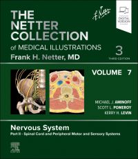
The Netter Collection of Medical Illustrations: Nervous System, Volume 7, Part II - Spinal Cord and Peripheral Motor and Sensory Systems, 3rd Edition
Author :
Edited by Michael J. Aminoff, MD, DSc, FRCP, Scott Pomeroy, MD, PhD and Kerry H. Levin, MD
Offering a concise, highly visual approach to the basic science and clinical pathology of the nervous system, this updated volume in The Netter Collection of Medical Illustrations (the CIBA "Green Books") contains unparalleled didactic illu
...view more
Offering a concise, highly visual approach to the basic science and clinical pathology of the nervous system, this updated volume in The Netter Collection of Medical Illustrations (the CIBA "Green Books") contains unparalleled didactic illustrations reflecting the latest medical knowledge. Revised by Drs. Michael J. Aminoff, Scott L. Pomeroy, and Kerry H. Levin, Spinal Cord and Peripheral Motor and Sensory Systems, Part 2 of the Nervous System, Volume 7, integrates core concepts of anatomy, physiology, and other basic sciences with common clinical correlates across health, medical, and surgical disciplines. Classic Netter art, updated and new illustrations, and modern imaging continue to bring medical concepts to life and make this timeless work an essential resource for students, clinicians, and educators.
Offering a concise, highly visual approach to the basic science and clinical pathology of the nervous system, this updated volume in The Netter Collection of Medical Illustrations (the CIBA "Green Books") contains unparalleled didactic illustrations reflecting the latest medical knowledge. Revised by Drs. Michael J. Aminoff, Scott L. Pomeroy, and Kerry H. Levin, Spinal Cord and Peripheral Motor and Sensory Systems, Part 2 of the Nervous System, Volume 7,integrates core concepts of anatomy, physiology, and other basic sciences with common clinical correlates across health, medical, and surgical disciplines. Classic Netter art, updated and new illustrations, and modern imaging continue to bring medical concepts to life and make this timeless work an essential resource for students, clinicians, and educators.
Key Features
Provides a highly visual overview of the anatomy, pathology, and major clinical syndromes of the nervous system, from cranial nerves and neuro-ophthalmology to spinal cord, neuropathies, autonomic nervous system, pain physiology, and neuromuscular disorders
Provides a concise overview of complex information by integrating anatomical and physiological concepts with clinical scenarios
Shares the experience and knowledge of Drs. Michael J. Aminoff, Scott L. Pomeroy, and Kerry H. Levin, and other experts at leading neurology centers
Compiles Dr. Frank H. Netter’s master medical artistry—an aesthetic tribute and source of inspiration for medical professionals for over half a century—along with new art in the Netter tradition for each of the major body systems, making this volume a powerful and memorable tool for building foundational knowledge and educating patients or staff
NEW! An eBook version is included with purchase. The eBook allows you to access all of the text, figures, and references, with the ability to search, make notes and highlights, and have content read aloud
Author Information
Edited by Michael J. Aminoff, MD, DSc, FRCP, Distinguished Professor Emeritus in Neurology; University of California San Francisco, USA; Scott Pomeroy, MD, PhD, Bronson Crothers Professor, Department of Neurology, Harvard Medical School; Consultant, Pediatric Oncology, Dana-Farber Cancer Institute; Neurologist-in-Chief, Department of Neurology, Boston Children's Hospital, Boston, Massachusetts, USA and Kerry H. Levin, MD, Chair, Department of Neurology, Director of the Neuromuscular Center at the Neurological Institute, Cleveland Clinic, Cleveland, OH, USA
Edited by Michael J. Aminoff, MD, DSc, FRCP, Distinguished Professor Emeritus in Neurology; University of California San Francisco, USA; Scott Pomeroy, MD, PhD, Bronson Crothers Professor, Department of Neurology, Harvard Medical School; Consultant, Pediatric Oncology, Dana-Farber Cancer Institute; Neurologist-in-Chief, Department of Neurology, Boston Children's Hospital, Boston, Massachusetts, USA and Kerry H. Levin, MD, Chair, Department of Neurology, Director of the Neuromuscular Center at the Neurological Institute, Cleveland Clinic, Cleveland, OH, USA
SECTION 1 CRANIAL NERVE AND NEURO-OPHTHALMOLOGIC DISORDERS Overview of Cranial Nerves 1.1 Distribution of Motor and Sensory Fibers 1.2 Overview of Cranial Nerves 1.3 Nerves and Nuclei Viewed in Phantom From Behind 1.4 Nerves and Nuclei in Lateral Dissection Cranial Nerve I: Olfactory Nerve 1.5 Olfactory Pathways 1.6 Olfactory Receptors 1.7 Olfactory Bulb and Nerve Cranial Nerve II: Optic Nerve 1.8 Eye 1.9 Cranial Nerve II and Visual Pathways 1.10 Optic Nerve Appearance 1.11 Retinal Projections to Thalamus, Midbrain, and Brainstem 1.12 Pupillary Light Reflex and the Accommodation Reflex Cranial Nerves III, IV, and VI (Oculomotor, Trochlear, and Abducens) 1.13 Oculomotor (III), Trochlear (IV), and Abducens Nerves (VI) 1.14 Nerves of Orbit and Cavernous Sinus 1.15 Damage to Cranial Nerve III 1.16 Control of Eye Movements 1.17 Control of Eye Movements: Pathology 1.18 Control of Eye Movements: Pathology (Continued) 1.19 Autonomic Innervation of the Eye Cranial Nerve V: Trigeminal Nerve 1.20 Trigeminal Nerve (V) 1.21 Trigeminal Nuclei: Afferent and Central Connections 1.22 Trigeminal Nuclei: Central and Peripheral Connections 1.23 Ophthalmic (V1) and Maxillary (V2) Nerves 1.24 Mandibular Nerve (V3) 1.25 Trigeminal Nerve Disorders Cranial Nerve VII: Facial Nerve 1.26 Pons: Level of the Genu of the Facial Nerve 1.27 Facial Nerve (VII) 1.28 Muscles of Facial Expression: Lateral View 1.29 Central Versus Peripheral Facial Paralysis 1.30 Facial Palsy Taste Receptors and Pathways 1.31 Anatomy of Taste Buds and Their Receptors 1.32 Tongue Cranial Nerve VIII: Vestibulocochlear Nerve 1.33 Vestibulocochlear Nerve (VIII) 1.34 Pathway of Sound Reception 1.35 Pathologic Causes of Vertigo 1.36 Canalith Repositioning (Epley Maneuver) 1.37 Afferent Auditory Pathways 1.38 Centrifugal Auditory Pathways 1.39 Vestibular Receptors 1.40 Cochlear Receptors Cranial Nerve IX: Glossopharyngeal Nerve and Otic Ganglion 1.41 Glossopharyngeal Nerve (IX) 1.42 Otic Ganglion Cranial Nerve X: Vagus Nerve 1.43 Vagus Nerve (X) 1.44 Motor and Sensory Branches From the Vagus Nerve 1.45 Neurogenic Disorders of Mouth and Pharynx (X and XII) Cranial Nerve XI: Accessory Nerve 1.46 Accessory Nerve (XI) 1.47 Clinical Findings in Cranial Nerve XI Damage Cranial Nerve XII: Hypoglossal Nerve 1.48 Hypoglossal Nerve Intermedullary Course 1.49 Hypoglossal Nerve (XII) 1.50 Disorders of Hypoglossal Nucleus and Nerve
SECTION 2 SPINAL CORD: ANATOMY AND MYELOPATHIES 2.1 Spinal Cord 2.2 Spinal Membranes and Nerve Roots 2.3 Arteries of Spinal Cord and Nerve Roots 2.4 Arteries of Spinal Cord: Intrinsic Distribution 2.5 Veins of Spinal Cord, Nerve Roots, and Vertebrae 2.6 Principal Fiber Tracts of Spinal Cord 2.7 Somesthetic System of Body 2.8 Corticospinal (Pyramidal) System: Motor Component 2.9 Rubrospinal Tract 2.10 Vestibulospinal Tracts 2.11 Reticulospinal and Corticoreticular Pathways 2.12 Spinal Origin or Termination of Major Descending Tracts and Ascending Pathways 2.13 Cytoarchitecture of Spinal Cord Gray Matter 2.14 Spinal Effector Mechanisms 2.15 Spinal Reflex Pathways 2.16 Spinal Cord Dysfunction 2.17 Sensory Impairment Related to Level of Spinal Cord Injury 2.18 Incomplete Spinal Cord Syndromes 2.19 Acute Spinal Cord Syndromes 2.20 Acute Spinal Cord Syndromes: Pathology, Etiology, and Diagnosis 2.21 Spinal Tumors 2.22 Extramedullary and Intramedullary Spinal Cord Tumors 2.23 Neuroimaging (MRI) Characteristics of Spinal Tumors 2.24 Syringomyelia 2.25 Subacute Combined Degeneration 2.26 Spinal Dural Fistulas and Arteriovenous Malformations 2.27 Cervical Spondylosis 2.28 Cervical Disk Herniation Causing Cord Compression 2.29 Infectious and Hereditary Myelopathies
SECTION 3 SPINAL TRAUMA 3.1 Spinal Column 3.2 Atlas and Axis 3.3 Cervical Vertebrae 3.4 External Craniocervical Ligaments 3.5 Internal Craniocervical Ligaments 3.6 Thoracic Vertebrae 3.7 Lumbar Vertebrae and Intervertebral Disk 3.8 Sacrum and Coccyx 3.9 Ligaments of Sacrum and Coccyx 3.10 Biomechanics of Spine and Spinal Cord Injuries: Distractive Flexion 3.11 Biomechanics of Spine and Spinal Cord Injuries: Compressive Flexion 3.12 Biomechanics of Spine and Spinal Cord Injuries: Distractive Extension 3.13 Cervical Spine Injury: Prehospital, Emergency Department, and Acute Management 3.14 Traction and Bracing 3.15 Anterior Cervical Spine Decompression and Stabilization 3.16 Posterior Cervical Stabilization and Fusion 3.17 Spinal Cord Injury Medical Issues
SECTION 4 NERVE ROOTS AND PLEXUS DISORDERS 4.1 Cervical Disk Herniation 4.2 Radiographic Diagnosis of Radiculopathy 4.3 Back Pain and Lumbar Disk Disease 4.4 Lumbar Disk Herniation: Clinical Manifestations 4.5 L4–5 Disk Extrusion 4.6 Lumbosacral Spinal Stenosis 4.7 Spinal Nerves 4.8 Dermal Segmentation 4.9 Thoracic Nerves 4.10 Thoracic Spinal Nerve Root Disorders 4.11 Diabetic Lumbosacral Radiculoplexus Neuropathy 4.12 Lumbar, Sacral, and Coccygeal Plexuses 4.13 Brachial Plexus 4.14 Brachial Plexus and Cervical Nerve Root Injuries at Birth 4.15 Brachial Plexopathy 4.16 Lumbosacral Plexopathy 4.17 Cervical Plexus
SECTION 5 MONONEUROPATHIES 5.1 Compression Neuropathies 5.2 Chronic Nerve Compression 5.3 Evaluation of Mononeuropathies 5.4 Radiologic Studies in Compression Neuropathy 5.5 Proximal Nerves of the Upper Extremity 5.6 Suprascapular and Musculocutaneous Nerves 5.7 Median Nerve 5.8 Proximal Median Neuropathies 5.9 Distal Median Neuropathies: Carpal Tunnel Syndrome 5.10 Distal Median Neuropathies: Carpal Tunnel Syndrome (Continued) 5.11 Ulnar Nerve 5.12 Ulnar Mononeuropathies: Potential Entrapment Sites 5.13 Radial Nerve 5.14 Radial Nerve Compression and Entrapment Neuropathies 5.15 Femoral and Lateral Femoral Cutaneous Nerves 5.16 Iliohypogastric, Ilioinguinal, Genitofemoral, and Obturator Nerves 5.17 Sciatic and Gluteal Nerves 5.18 Sciatic and Posterior Femoral Cutaneous Nerves 5.19 Fibular (Peroneal) and Tibial Nerves 5.20 Peroneal Nerve Compression 5.21 Tibial Nerve 5.22 Dermatomal and Cutaneous Nerve Patterns 5.23 Dermatomes
SECTION 6 PERIPHERAL NEUROPATHIES 6.1 Peripheral Nerve 6.2 Histology of Peripheral Nerve 6.3 Cell Types of Nervous System 6.4 Resting Membrane Potential 6.5 Ion Channel Mechanics and Action Potential Generation 6.6 Neurophysiology and Peripheral Nerve Demyelination 6.7 Impulse Propagation 6.8 Conduction Velocity 6.9 Visceral Efferent Endings 6.10 Cutaneous Receptors 6.11 Pacinian Corpuscle 6.12 Muscle and Joint Receptors 6.13 Proprioceptive Reflex Control of Muscle Tension 6.14 Charcot-Marie-Tooth Disease Overview (Hereditary Motor and Sensory Neuropathy) 6.15 Charcot-Marie-Tooth Disease: Common Types 6.16 Early Onset and Other Rare Forms of Charcot-Marie-Tooth Disease and Inherited Neuropathies 6.17 Hereditary Sensory and Autonomic Neuropathy 6.18 Guillain-Barré Syndrome 6.19 Guillain-Barré Syndrome (Continued) 6.20 Chronic Inflammatory Demyelinating Polyradiculoneuropathy 6.21 Diabetic Neuropathies 6.22 Monoclonal Protein–Associated Neuropathies 6.23 Distal Acquired Demyelinating Symmetric (DADS) Neuropathy 6.24 Vasculitic Neuropathy and Other Connective Tissue Disorders Associated With Neuropathy 6.25 Sjögren Syndrome 6.26 Immunopathogenesis of Guillain-Barré Syndrome 6.27 Peripheral Neuropathy Caused by Heavy Metal Poisoning 6.28 Metabolic, Toxic, and Nutritional Peripheral Neuropathies 6.29 Leprosy and Other Infections Sometimes Causing Peripheral Neuropathy
SECTION 7 AUTONOMIC NERVOUS SYSTEM AND ITS DISORDERS 7.1 General Topography of Autonomic Nervous System 7.2 General Topography of Autonomic Nervous System (Continued) 7.3 Autonomic Reflex Pathways 7.4 Cholinergic and Adrenergic Nerves 7.5 Autonomic Nerves in Head and Neck 7.6 Autonomic Nerves in Neck 7.7 Autonomic Innervation of Eye 7.8 Autonomic Innervation of Eye (Continued) 7.9 Autonomic Nerves in Thorax 7.10 Innervation of Heart 7.11 Innervation of Blood Vessels 7.12 Carotid Body and Carotid Sinus 7.13 Autonomic Nerves and Ganglia in Abdomen 7.14 Innervation of Stomach and Proximal Duodenum 7.15 Innervation of Intestines 7.16 Autonomic Innervation of Small Intestine 7.17 Enteric Plexuses 7.18 Innervation of Liver and Biliary Tract 7.19 Innervation of Adrenal Glands 7.20 Autonomic Nerves and Ganglia in Pelvis 7.21 Innervation of Kidneys, Ureters, and Urinary Bladder 7.22 Innervation of Urinary Bladder and Lower Ureter 7.23 Innervation of Reproductive Organs 7.24 Female Reproductive Organs 7.25 Autonomic Testing 7.26 Cardiovagal Testing and Head-up Tilting 7.27 Abnormal Pupillary Conditions 7.28 Clinical Presentation of Autonomic Disorders 7.29 Multiple System Atrophy
SECTION 8 PAIN 8.1 Neuroanatomy of the Ascending Pain Pathways 8.2 Neuroanatomy of the Ascending Pain Pathways (Continued) 8.3 Descending Nociceptive Pathways and Neurochemical Foundations of Descending Pain Modulation 8.4 Endorphin System 8.5 Nociceptive Processing and Central Nervous System Correlates of Pain 8.6 Central Nervous System Neurotransmitters, Receptors, and Drug Targets 8.7 Thalamic Pain Syndrome 8.8 Clinical Manifestations Related to Thalamus Site in Intracerebral Hemorrhage 8.9 Complex Regional Pain 8.10 Herpes Zoster 8.11 Occipital Neuralgia 8.12 Myofascial Factors in Low Back Pain 8.13 Posterior Abdominal Wall: Internal View 8.14 Lumbar Zygapophyseal Joint Back Pain 8.15 Low Back Pain and Effects of Lumbar Hyperlordosis and Flexion on Spinal Nerves 8.16 Examination of the Patient With Low Back Pain 8.17 Osteoporosis 8.18 Diagnosis of Low Back, Buttock, and Hip Pain 8.19 Hip Joint Involvement in Osteoarthritis 8.20 Painful Peripheral Neuropathy 8.21 Peripheral Neuropathies: Clinical Manifestations 8.22 Functional Neurologic Disorders 8.23 Somatoform Conversion Reactions
SECTION 10 MOTOR NEURON AND ITS DISORDERS 10.1 Peripheral Nervous System: Overview 10.2 Spinal Cord and Neuronal Cell Body With Motor, Sensory, and Autonomic Components of the Peripheral Nerve 10.3 Motor Unit 10.4 Motor Unit Potentials 10.5 Primary Motor Neuron Disease 10.6 Amyotrophic Lateral Sclerosis 10.7 Clinical Manifestations of Amyotrophic Lateral Sclerosis (Continued) 10.8 Mimics of Amyotrophic Lateral Sclerosis 10.9 Diagnosis of Amyotrophic Lateral Sclerosis 10.10 Treatment of Amyotrophic Lateral Sclerosis 10.11 Spinal Muscular Atrophy and Spinal Bulbar Muscular Atrophy
SECTION 11 NEUROMUSCULAR JUNCTION AND ITS DISORDERS 11.1 Neuromuscular Junction 11.2 Physiology of Neuromuscular Junction 11.3 Synaptic Transmission 11.4 Synaptic Transmission (Continued) 11.5 Repetitive Motor Nerve Stimulation 11.6 Patient With Lambert-Eaton Myasthenic Syndrome 11.7 Myasthenia Gravis 11.8 Myasthenia Gravis (Continued) 11.9 Immunopathology of Myasthenia Gravis 11.10 Presynaptic Neuromuscular Junction Transmission Disorders: Lambert-Eaton Myasthenic Syndrome and Infantile Botulism 11.11 Congenital Myasthenic Syndromes 11.12 Foodborne Neurotoxins
SECTION 12 MUSCLE AND ITS DISORDERS 12.1 Muscle Fiber Anatomy: Basic Sarcomere Subdivisions 12.2 Muscle Fiber Anatomy: Biochemical Mechanics of Contraction 12.3 Muscle Membrane, T Tubules, and Sarcoplasmic Reticulum 12.4 Muscle Response to Nerve Stimulation 12.5 Metabolism of Muscle Cell 12.6 Muscle Fiber Types 12.7 Overview of Myopathies: Clinical Approach 12.8 Dystrophinopathies 12.9 Duchenne Muscular Dystrophy 12.10 Dystrophinopathies (Continued) 12.11 Myotonic Dystrophy and Other Myotonic Disorders 12.12 Myotonic Dystrophy and Other Myotonic Disorders (Continued) 12.13 Other Types of Muscular Dystrophy 12.14 Polymyositis and Dermatomyositis 12.15 Polymyositis and Dermatomyositis (Continued) 12.16 Inclusion Body Myositis 12.17 Immunopathogenesis of Inflammatory Myopathies 12.18 Endocrine, Toxic, and Critical Illness Myopathies 12.19 Myopathies: Hypokalemia/Hyperkalemia and Periodic Paralyses Channelopathies 12.20 Metabolic and Mitochondrial Myopathies 12.21 Myoglobinuric Syndromes Including Malignant Hyperthermia
https://www.elsevierhealth.com.au/the-netter-collection-of-medical-illustrations-nervous-system-volume-7-part-ii-spinal-cord-and-peripheral-motor-and-sensory-systems-9780323880855.html310884The Netter Collection of Medical Illustrations: Nervous System, Volume 7, Part II - Spinal Cord and Peripheral Motor and Sensory Systemshttps://www.elsevierhealth.com.au/media/catalog/product/9/7/9780323880855_2.jpg116.96155.95AUDInStock/Medicine/Anatomy/Medicine/Anatomy/Books/Books/Japan Titles/Medicine & Surgery/Anatomy/Books/Medical Students/Medical education/Medical Students/Medical education/Students/Anatomy and physiology/Students/First years505461552599025054664525503952631604335138438872254964465496447505802650580251488653650545535145120525983341826924335088530301953030185058024Offering a <b>concise, highly visual approach </b>to the basic science and clinical pathology of the nervous system, this updated volume in <i>The Netter Collection of Medical Illustrations</i> (the CIBA "Green Books") contains unparalleled didactic illustrations reflecting the latest medical knowledge. Revised by Drs. Michael J. Aminoff, Scott L. Pomeroy, and Kerry H. Levin, <b>Spinal Cord and Peripheral Motor and Sensory Systems, Part 2 of the Nervous System, Volume 7</b><i>,</i> <b>integrates core concepts of anatomy, physiology, and other basic sciences with common clinical correlates</b> across health, medical, and surgical disciplines. <b>Classic Netter art, updated and new illustrations, and modern imaging</b> continue to bring medical concepts to life and make this timeless work an essential resource for students, clinicians, and educators. Offering a <b>concise, highly visual approach </b>to the basic science and clinical pathology of the nervous system, this updated volume in <i>The Netter Collection of Medical Illustrations</i> (the CIBA "Green Books") contains unparalleled didactic illustrations reflecting the latest medical knowledge. Revised by Drs. Michael J. Aminoff, Scott L. Pomeroy, and Kerry H. Levin, <b>Spinal Cord and Peripheral Motor and Sensory Systems, Part 2 of the Nervous System, Volume 7</b><i>,</i> <b>integrates core concepts of anatomy, physiology, and other basic sciences with common clinical correlates</b> across health, medical, and surgical disciplines. <b>Classic Netter art, updated and new illustrations, and modern imaging</b> continue to bring medical concepts to life and make this timeless work an essential resource for students, clinicians, and educators.00add-to-cart97803238808552024ProfessionalEdited by Michael J. Aminoff, MD, DSc, FRCP, Scott Pomeroy, MD, PhD and Kerry H. Levin, MD20253BookOther268 illustrations (268 in full color)Elsevier30215 Apr 2024Available for Pre-OrderEdited by <STRONG>Michael J. Aminoff</STRONG>, MD, DSc, FRCP, Distinguished Professor Emeritus in Neurology; University of California San Francisco, USA; <STRONG>Scott Pomeroy</STRONG>, MD, PhD, Bronson Crothers Professor, Department of Neurology, Harvard Medical School; Consultant, Pediatric Oncology, Dana-Farber Cancer Institute; Neurologist-in-Chief, Department of Neurology, Boston Children's Hospital, Boston, Massachusetts, USA and <STRONG>Kerry H. Levin</STRONG>, MD, Chair, Department of Neurology, Director of the Neuromuscular Center at the Neurological Institute, Cleveland Clinic, Cleveland, OH, USABooksBookNetter Green Book CollectionUnited StatesYesYesNoNoPlease SelectPlease SelectPlease Select