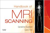
By Geraldine Burghart, MA, RT(R)(MR)(M) and Carol Ann Finn, RT(R)(MR)
Ensure high-quality diagnostic images with this practical scanning reference! Designed to help you plan and acquire MRI images, Handbook of MRI Scanning, by Geraldine Burghart and Carol Ann Finn, includes the step-by-step scanning protocols you need
...view more
Ensure high-quality diagnostic images with this practical scanning reference! Designed to help you plan and acquire MRI images, Handbook of MRI Scanning, by Geraldine Burghart and Carol Ann Finn, includes the step-by-step scanning protocols you need to produce optimal images. Coverage of all body regions prepares you to perform virtually any scan. Going beyond the referencing and recognition of three-plane, cross-sectional anatomy, each chapter demonstrates appropriate slice placements, typical midline images of each plane, and detailed line drawings of the pertinent anatomy corresponding to the midline images. With this handbook, you can conceptualize an entire scan and its intended outcome prior to performing the scan on a patient. Keep the book at your console -- it’s ideal for quick reference!
Ensure high-quality diagnostic images with this practical scanning reference! Designed to help you plan and acquire MRI images, Handbook of MRI Scanning, by Geraldine Burghart and Carol Ann Finn, includes the step-by-step scanning protocols you need to produce optimal images. Coverage of all body regions prepares you to perform virtually any scan. Going beyond the referencing and recognition of three-plane, cross-sectional anatomy, each chapter demonstrates appropriate slice placements, typical midline images of each plane, and detailed line drawings of the pertinent anatomy corresponding to the midline images. With this handbook, you can conceptualize an entire scan and its intended outcome prior to performing the scan on a patient. Keep the book at your console -- it’s ideal for quick reference!
Key Features
Consistent, clinically based layout of the sections makes scanning information easy to use with three images per page to demonstrate clinical sequences in MRI examinations.
Handy, pocket size offers easy, immediate access right at the console.
600 images provide multiple views and superb anatomic detail.
Suggested technical parameters are provided in convenient tables for quick reference with space to write in site-specific protocols or equipment variations.
Author Information
By Geraldine Burghart, MA, RT(R)(MR)(M), Associate Professor, Radiologic Technology Program, Department of Nursing and Allied Health Sciences, City University of New York, BCC, Bronx, New York and Carol Ann Finn, RT(R)(MR), Manager, MRI Department, New York Presbyterian Hospital, Columbia Campus, New York, New York
By Geraldine Burghart, MA, RT(R)(MR)(M), Associate Professor, Radiologic Technology Program, Department of Nursing and Allied Health Sciences, City University of New York, BCC, Bronx, New York and Carol Ann Finn, RT(R)(MR), Manager, MRI Department, New York Presbyterian Hospital, Columbia Campus, New York, New York
"This book provides technologists and students with not only a baseline for MR image acquisition, but also a standard of quality that should be consistently duplicated to provide the healthcare team with quality diagnostic images. This is a must read for any student or beginning technologist. It does a great job of providing a practical, realistic approach to MRI that avoids confusing physics and equations. I have never seen another book that packed as much useful and simplistic information into such a small package." - Karl Freund, RT(R) MR, Froedtert Hospital (4 Star Doody Rating)
https://www.elsevierhealth.com.au/handbook-of-mri-scanning-9780323068185.html293726Handbook of MRI Scanninghttps://www.elsevierhealth.com.au/media/catalog/product/9/7/9780323068185_7.jpg98.05108.94AUDInStock/Health Professions/Radiography/Books/Health Professions/Radiography/Books/Health Professions/Radiography/Japan Titles/Books43351204388722505458852550395259870526316050546641418269243350924886536505455851451205259838<P>Ensure high-quality diagnostic images with this practical scanning reference! Designed to help you plan and acquire MRI images, <b>Handbook of MRI Scanning</b>, by Geraldine Burghart and Carol Ann Finn, includes the step-by-step scanning protocols you need to produce optimal images. Coverage of all body regions prepares you to perform virtually any scan. Going beyond the referencing and recognition of three-plane, cross-sectional anatomy, each chapter demonstrates appropriate slice placements, typical midline images of each plane, and detailed line drawings of the pertinent anatomy corresponding to the midline images. With this handbook, you can conceptualize an entire scan and its intended outcome prior to performing the scan on a patient. Keep the book at your console -- it’s ideal for quick reference!</P> <P>Ensure high-quality diagnostic images with this practical scanning reference! Designed to help you plan and acquire MRI images, <b>Handbook of MRI Scanning</b>, by Geraldine Burghart and Carol Ann Finn, includes the step-by-step scanning protocols you need to produce optimal images. Coverage of all body regions prepares you to perform virtually any scan. Going beyond the referencing and recognition of three-plane, cross-sectional anatomy, each chapter demonstrates appropriate slice placements, typical midline images of each plane, and detailed line drawings of the pertinent anatomy corresponding to the midline images. With this handbook, you can conceptualize an entire scan and its intended outcome prior to performing the scan on a patient. Keep the book at your console -- it’s ideal for quick reference!</P>00add-to-cart97803230681852011 and earlierProfessionalBy Geraldine Burghart, MA, RT(R)(MR)(M) and Carol Ann Finn, RT(R)(MR)20111Book152w x 229h (6.00" x 9.00")Approx. 422 illustrationsMosby39630 Dec 2010Please allow 3-4 weeks for deliveryBy <STRONG>Geraldine Burghart</STRONG>, MA, RT(R)(MR)(M), Associate Professor, Radiologic Technology Program, Department of Nursing and Allied Health Sciences, City University of New York, BCC, Bronx, New York and <STRONG>Carol Ann Finn</STRONG>, RT(R)(MR), Manager, MRI Department, New York Presbyterian Hospital, Columbia Campus, New York, New YorkBooksBookUnited StatesNoNoNoNoPlease SelectPlease SelectPlease Select