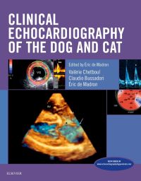
Clinical Echocardiography of the Dog and Cat, 1st Edition
Author :
By Eric DURRIEU DE MADRON, Valérie Chetboul, Valérie Chetboul est docteur vétérinaire, agrégée de pathologie médicale et diplômée de l'European College of Veterinary Internal Medicine. and Claudio Bussadori
Covering both classical modalities of echocardiography and newer techniques, Clinical Echocardiography of the Dog and Cat shows how to assess, diagnose, and treat canine and feline heart disease. A clinical approach demonstrates how these modalities
...view more
Covering both classical modalities of echocardiography and newer techniques, Clinical Echocardiography of the Dog and Cat shows how to assess, diagnose, and treat canine and feline heart disease. A clinical approach demonstrates how these modalities may be used to acquire images, and then how you can recognize and identify patterns, relate them to different diseases, and manage patient care with those findings. The print book includes a companion website with 50 videos of cardiac ultrasound exams and procedures. Written by veterinary cardiology specialists and echocardiographers Eric de Madron, Valerie Chetboul, and Claudio Bussadori, this indispensable echocardiology resource is ideal for general practitioner veterinarians as well as specialists, including cardiologists and radiologists.
Covering both classical modalities of echocardiography and newer techniques, Clinical Echocardiography of the Dog and Cat shows how to assess, diagnose, and treat canine and feline heart disease.A clinical approach demonstrateshow these modalities may be used to acquire images, and then how you can recognize and identify patterns, relate them to different diseases, and manage patient care with those findings. The print book includes a companion website with 50 videos of cardiac ultrasound exams and procedures. Written by veterinary cardiology specialists and echocardiographers Eric de Madron, Valerie Chetboul, and Claudio Bussadori, this indispensable echocardiology resource is ideal for general practitioner veterinarians as well as specialists, including cardiologists and radiologists.
Key Features
Dedicated coverage of canine and feline echocardiography
emphasizes a more in-depth discussion of cardiac ultrasound, including the newest ones such as Tissue Doppler and speckle tracking imaging, and transesophageal and 3D echocardiography.
A practical, clinical approach shows how these echocardiographic modalities are not just research tools, but useful in diagnosing and staging heart disease in day-to-day practice.
Book plus website consolidates offers current information into a single cohesive source covering classical modalities and newer techniques, as well as updates relating to normal echocardiographic examinations and values.
50 videos on the companion website demonstrate how to perform echocardiography procedures, illustrating points such as swirling volutes, color flow display of blood flows, dynamic collapses secondary to pericardial effusion, and tumors flicking in and out of the echocardiographic field.
A section on presurgical assessment helps you assess risk and prepare for catheter-based correction of cardiac defects — accurate measurements and proper device selection are key to a successful procedure.
Over 400 full-color illustrations and 42 summary tables help you achieve precise, high-quality imaging for accurate assessment, including photographs of cadaver animal specimens to clarify the relationship between actual tissues in health and disease and their images.
Author Information
By Eric DURRIEU DE MADRON, Docteur vétérinaire DVM, DACVIM (cardiologie), DECVIM (Médecine interne) au Canada; Valérie Chetboul, Valérie Chetboul est docteur vétérinaire, agrégée de pathologie médicale et diplômée de l'European College of Veterinary Internal Medicine., Vétérinaire, unité de Cardiologie, centre hospitalier vétérinaire d'Alfort, Maisons-Alfort, France. and Claudio Bussadori
By Eric DURRIEU DE MADRON, Docteur vétérinaire DVM, DACVIM (cardiologie), DECVIM (Médecine interne) au Canada; Valérie Chetboul, Valérie Chetboul est docteur vétérinaire, agrégée de pathologie médicale et diplômée de l'European College of Veterinary Internal Medicine., Vétérinaire, unité de Cardiologie, centre hospitalier vétérinaire d'Alfort, Maisons-Alfort, France. and Claudio Bussadori
PART I: NORMAL ECHOCARDIOGRAPHIC EXAMINATION Chapter 1: Normal Views: 2D, TM, Spectral and Color Doppler Modes Eric de Madron • The different modes • General considerations • Normal bi-dimensional views • Normal spectral Doppler examination • Color Doppler Chapter 2: Normal Echocardiographic Values: 2D, TM and Spectral Doppler Modes Eric de Madron • Normal TM and 2D values • Normal spectral Doppler values Chapter 3: Intraoperator and Interoperator Variability Valerie Chetboul • Repeatability, reproducibility and operator performance • Operator effect on the variability of echocardiographic measurements • Practical implications PART II: NEW TECHNIQUES OF ULTRASONOGRAPHIC CARDIAC IMAGING Chapter 4: Myocardial Tissue Doppler, Derived Techniques and Speckle Tracking Imaging Valerie Chetboul • Normal myocardial kinetics • Myocardial Tissue Doppler or Tissue Doppler imaging (TDI) • Derived techniques from TDI: tissue tracking imaging, strain and strain rate imaging • Speckle tracking imaging Chapter 5: Trans-Esophageal Echocardiography Claudio Bussadori • Introduction and technical aspects • Considerations with respect to the echocardiographer • ETO examination protocol • Indications and applications of ETO Chapter 6: Veterinary Applications of Three-Dimensional Echocardiography Claudio Bussadori • 3D echocardiography technology • The different modes of 3D echocardiography • 3D echocardiography in veterinary medicine • Patent ductus arteriosus • Pulmonic valve valvuloplasty PART III: HEMODYNAMIC EVALUATION Chapter 7: Global Left Ventricular Systolic Function Evaluation Eric de Madron • Left ventricular anatomy • Radial systolic function: shortening fraction • Systolic indices derived from ventricular volumes • Longitudinal systolic function evaluation • Spectral Doppler indices: systolic time intervals and index of myocardial performance (Tei index) Chapter 8: Diastolic Function Evaluation Eric de Madron • Phases of diastole and its determinants • Types of diastolic dysfunctions • Echocardiographic indices of diastolic function Chapter 9: Echocardiographic Assessment of the Left Ventricular Filling Pressure Eric de Madron • Left atrial pressure curve • Echocardiographic indices of the left ventricular filling pressure • Other echocardiographic criteria Chapter 10: Right Ventricular Systolic Function Evaluation Eric de Madron • Radial right ventricular systolic function: right ventricular shortening fractions • Longitudinal right ventricular systolic function assessment • Systolic time intervals and index of myocardial performance (Tei index) • Spectral Doppler indices PART IV: ECHOCARDIOGRAPHY OF ACQUIRED HEART DISEASES Chapter 11: Evaluation and Quantification of Acquired Valvular Regurgitations in the Dog Eric de Madron • Mitral regurgitation • Tricuspid regurgitation • Aortic regurgitation • Pulmonic regurgitation Chapter 12: Canine Dilated Cardiomyopathy and Other Cardiomyopathies Valerie Chetboul • Primary dilated cardiomyopathy • Other cardiomyopathies Chapter 13: Feline Cardiomyopathies Assessment Eric de Madron • Hypertrophic cardiomyopathies • Restrictive cardiomyopaties • Dilated cardiomyopathies • Atypical cardiomyopathies • Thrombotic risk evaluation Chapter 14: Pulmonary Hypertension Valerie Chetboul • Review of pulmonary arterial hypertension (PAH) • Conventional echocardiography and PAH • Conventional Doppler and PAH • New imaging modalities and PAH Chapter 15: Dirofilariosis: Specific Bi-Dimensional Findings Eric de Madron • Echocardiographic characteristics of dirofilaria • Other echocardiographic anomalies associated with dirofilariosis Chapter 16: Cardiac Manifestations of Systemic Diseases Valerie Chetboul, Eric de Madron • Systemic arterial hypertension • Hyperthyroid cardiopathy • Anemia Chapter 17: Pericardial Diseases Eric de Madron • Pericardial anatomy and function • Pericardial effusions • Constrictive pericarditis • Congenital pericardial defects Chapter 18: Cardiac Tumors Claudio Bussadori • Types of tumors and prevalence • Goals of echocardiography in the assessment of cardiac tumors • Specific echocardiographic presentation PART V: CONGENITAL CARDIOPATHIES Chapter 19: Congenital Cardiopathies Claudio Bussadori • Aortic stenosis • Pulmonic stenosis • Patent ductus arteriosus • Ventricular septal defects • Atrial septal defects • Mitral valve dysplasia • Tricuspid valve dysplasia • Tetralogy of Fallot • Cor triatriatum • Endocardial cushion defects Chapter 20: Echocardiographic Assessment of Congenital Cardiopathies Before and After Intervention Claudio Bussadori • Valvular pulmonic stenosis • Patent ductus arteriosus • Ventricular septum defects • Atrial septal defects
https://www.elsevierhealth.com.au/clinical-echocardiography-of-the-dog-and-cat-9780323316507.html294131Clinical Echocardiography of the Dog and Cathttps://www.elsevierhealth.com.au/media/catalog/product/9/7/9780323316507_7.jpg223.47297.95AUDInStock/Veterinary/Small Animal/Books/Veterinary/Small Animal/Books/Books/Veterinary/Small Animal/Japan Titles43351274388722505460450546645255039525988652631601418269243350964886536505456251451205259842<P>Covering both classical modalities of echocardiography and newer techniques, <B>Clinical Echocardiography of the Dog and Cat </B>shows how to assess, diagnose, and treat canine and feline heart disease.<B> </B>A clinical approach demonstrates<B> </B>how these modalities may be used to acquire images, and then how you can recognize and identify patterns, relate them to different diseases, and manage patient care with those findings. The print book includes a companion website with 50 videos of <b>cardiac ultrasound exams and </b>procedures. Written by veterinary cardiology specialists and echocardiographers Eric de Madron, Valerie Chetboul, and Claudio Bussadori, this indispensable echocardiology resource is ideal for general practitioner veterinarians as well as specialists, including cardiologists and radiologists.</P> <P>Covering both classical modalities of echocardiography and newer techniques, <B>Clinical Echocardiography of the Dog and Cat </B>shows how to assess, diagnose, and treat canine and feline heart disease.<B> </B>A clinical approach demonstrates<B> </B>how these modalities may be used to acquire images, and then how you can recognize and identify patterns, relate them to different diseases, and manage patient care with those findings. The print book includes a companion website with 50 videos of <b>cardiac ultrasound exams and </b>procedures. Written by veterinary cardiology specialists and echocardiographers Eric de Madron, Valerie Chetboul, and Claudio Bussadori, this indispensable echocardiology resource is ideal for general practitioner veterinarians as well as specialists, including cardiologists and radiologists.</P>00add-to-cart97803233165072015ProfessionalBy Eric DURRIEU DE MADRON, Valérie Chetboul, Valérie Chetboul est docteur vétérinaire, agrégée de pathologie médicale et diplômée de l'European College of Veterinary Internal Medicine. and Claudio Bussadori20151Book216w x 276h (8.50" x 10.875")Approx. 589 illustrations (542 in full color)Elsevier Masson3601 Jun 2015Please allow 3-4 weeks for deliveryBy <STRONG>Eric DURRIEU DE MADRON</STRONG>, Docteur vétérinaire DVM, DACVIM (cardiologie), DECVIM (Médecine interne) au Canada; <STRONG>Valérie Chetboul</STRONG>, Valérie Chetboul est docteur vétérinaire, agrégée de pathologie médicale et diplômée de l'European College of Veterinary Internal Medicine., Vétérinaire, unité de Cardiologie, centre hospitalier vétérinaire d'Alfort, Maisons-Alfort, France. and <STRONG>Claudio Bussadori</STRONG>BooksBookUnited StatesNoNoNoNoPlease SelectPlease SelectPlease Select